
Do White Matter Lesions Mean You Have Multiple Sclerosis or Dementia?
You just got your brain MRI results back, and they mention "white matter lesions" or "white spots", but your primary doctor's response left you with more questions than answers. What now?
If you're worried these findings suggest multiple sclerosis (MS) or dementia, you're not alone. Understanding what these findings mean for your specific situation is crucial.
The good news: Most white matter lesions are not due to MS or dementia, though some are, and here is how to know the difference and what to do next.
What Is White Matter, and How Is It Different from Gray Matter?
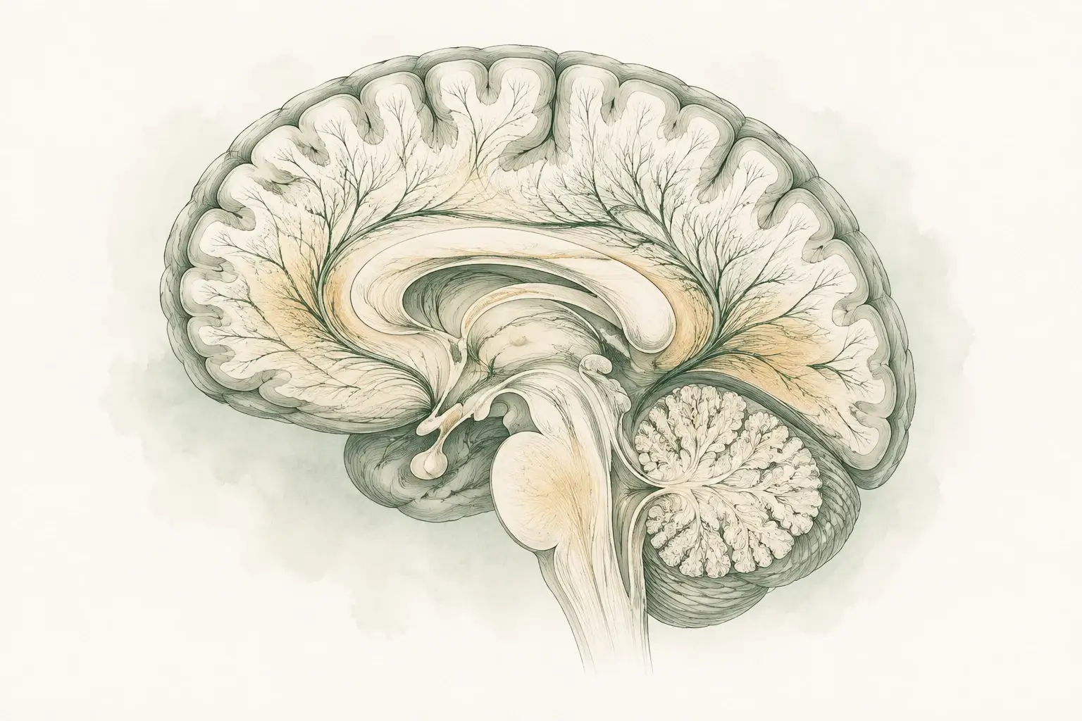
The brain's connecting tissue
- Connections: White matter carries signals between brain regions.
- MRI finding: Bright spots can reflect changes in this tissue.
- Context: Their meaning depends on the pattern and the clinical picture.
Before we dive deeper into white matter lesions, let's first clarify what white matter and gray matter actually are. Your brain is made up of two primary types of tissue, each with its own distinct yet interconnected role:
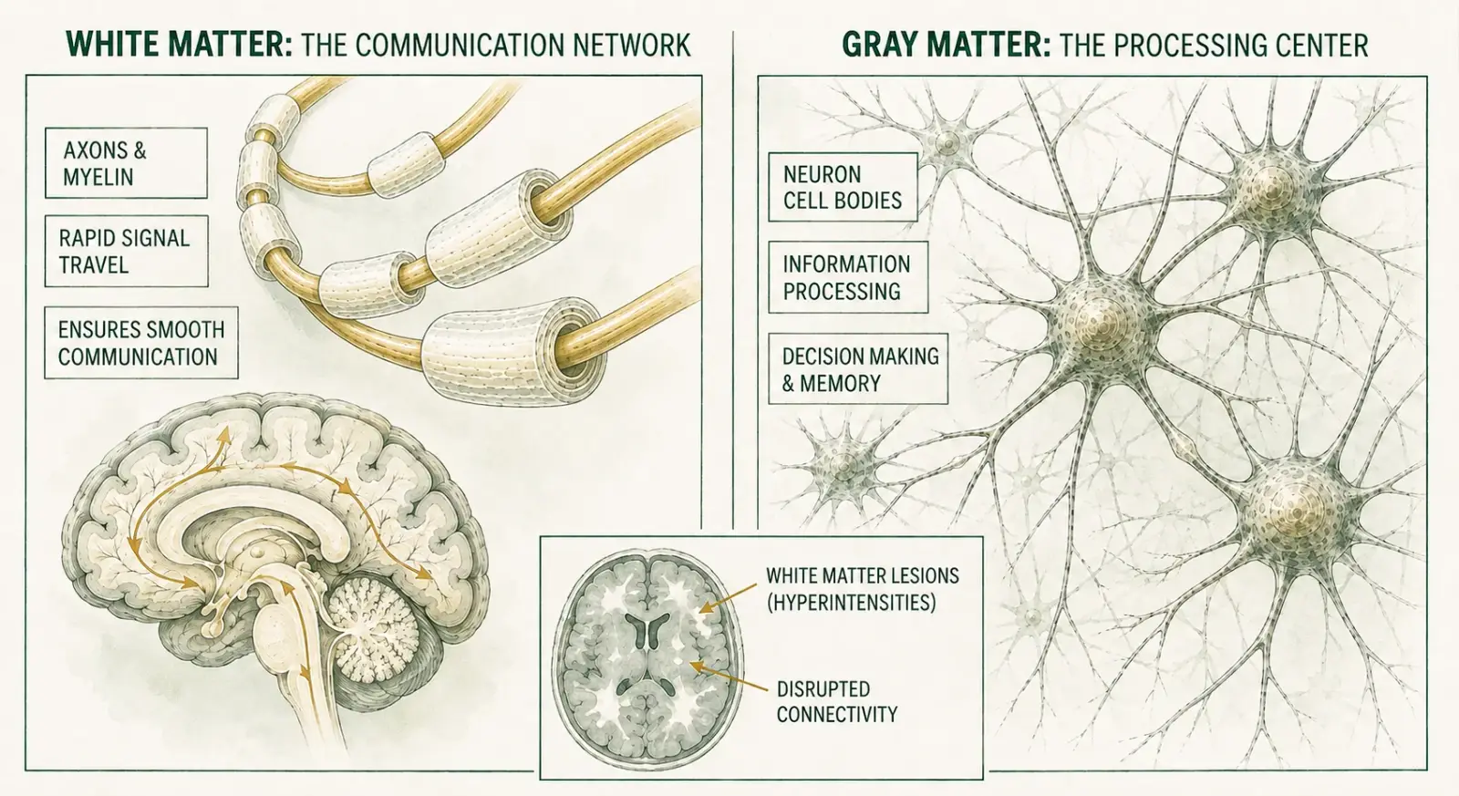
White Matter: The Brain's Communication Network
Made up of nerve fibers, called axons, and supporting cells
Covered by a fatty substance called myelin, which gives it a white color
Acts like insulated electrical wires, helping signals travel rapidly between different parts of the brain
Ensures smooth communication and coordination between regions of gray matter
Gray Matter: The Brain's Processing Center
Involved in processing and interpreting information
Called "gray matter" because of its darker color
Consists of neuron cell bodies, dendrites, and synapses (connections between neurons)
Responsible for processing sensory information, decision-making, emotional regulation, memory, and muscle control.
What Are White Matter Lesions?
White matter lesions refer to areas in the brain where the white matter tissue has experienced damage or changes. They signal that something has disrupted normal brain connectivity or blood flow in that area.
On MRI they show up as bright spots, and reports describe them in a few standard words, most often white matter hyperintensities or T2/FLAIR hyperintense foci.
What Do T2, FLAIR, and Hyperintense Foci Mean?
- T2 and FLAIR: two kinds of MRI image. T2 shows water as bright; FLAIR (fluid-attenuated inversion recovery) darkens the fluid around the brain so nearby spots stand out.
- Hyperintense: brighter than the tissue around it.
- Foci: spots; one is a focus. Punctate means tiny, scattered means spread apart.
- Nonspecific: the look does not point to one cause. It describes the image, not a diagnosis.
- Gliosis: scar-like tissue left by the brain's support cells after an earlier injury.
So "a few scattered T2/FLAIR hyperintense foci" means a few separate bright spots.
Other report words describe how much change there is and where it comes from. Most of them belong to the blood vessel pattern explained under white matter disease below:
- Confluent: spots that have merged into larger patches.
- Chronic small vessel ischemic changes, microangiopathy, leukoaraiosis: three names for the same finding, wear in the brain's smallest blood vessels.
- Fazekas 1, 2 or 3 (or mild, moderate, severe): a grade for how much of that change there is, from a few dots to large merged areas.
- "Not unusual for age": the amount of change is what the radiologist expects to see at your age.
These spots are very common, more so with age, and many are harmless. A few scattered ones often cause no symptoms. While they can sound alarming, these lesions vary greatly in significance, depending on their number, size, location, and the underlying cause.
What Causes White Matter Lesions In The Brain?
White matter lesions can result from a wide range of issues, some relatively benign and others potentially more serious. Common causes include:
Vascular Causes (related to blood vessels):
Aging: Normal aging can cause gradual wear-and-tear on small blood vessels, resulting in lesions.
High Blood Pressure: Chronic hypertension damages the tiny arteries supplying your brain.
Diabetes and High Cholesterol: These conditions affect blood vessels, leading to reduced blood flow and oxygen delivery.
Small Vessel Disease: A common cause where small blood vessels become damaged due to age or cardiovascular risks.
Smoking: Smoking can damage the blood vessels and lead to white matter lesions.
Non-Vascular Causes (not directly related to blood vessels):
Multiple Sclerosis (MS): Inflammatory diseases like MS can also lead to the formation of white matter lesions as a result of damage to the myelin sheath surrounding brain cells. Recognizing the early signs of MS can help you get evaluated before significant damage accumulates.
Demyelinating Disorders: Conditions that cause the loss of myelin, such as neuromyelitis optica or MOG disease, can result in white matter abnormalities.
Migraines: Chronic migraines can sometimes be associated with subtle white matter changes.
Infections: Certain viral infections (like HIV or Lyme disease) can affect white matter.
Toxic Substances: Chronic alcohol abuse, drug use (e.g., cocaine), or exposure to certain toxins can damage white matter.
Metabolic Issues: Deficiencies in nutrients like Vitamin B12 can lead to white matter abnormalities.
Genetic Conditions (Leukodystrophies): Rare inherited disorders affecting myelin formation.
Traumatic Brain Injury (TBI): A severe impact to the head can cause white matter lesions.
Tumors: Benign or malignant tumors can cause white matter lesions.
Hematological Disorders: Conditions like leukemia or lymphoma can lead to white matter changes.
Autoimmune Disorders: Conditions like lupus or rheumatoid arthritis can cause white matter lesions.
Depending on the type of underlying condition, white matter lesions may progress slowly or rapidly.
Hidden Risk Factors You Should Know
Smoking, poor diet and physical inactivity significantly increase your risk of white matter lesions. These lifestyle factors damage small blood vessels in your brain over time, creating a cumulative effect that accelerates brain aging.
White matter disease: when the spots come from small blood vessels
If your report says "white matter disease", it is usually describing the most common reason for white spots: wear in the brain's smallest blood vessels, not MS. Radiologists use several names for this one finding: white matter disease, chronic small vessel ischemic changes, microvascular ischemic disease, microangiopathy and leukoaraiosis. Over the years, that wear leaves small patches of white matter with less blood flow.
Radiologists sometimes write "demyelination" for these spots as well. It sounds like MS, but demyelination does not always mean MS, and here it means loss of myelin, the fibers' insulation, caused by the same blood vessel wear.
Reports may grade how much change there is, usually on the Fazekas scale from 0 to 3:
- 0: none.
- 1, mild: a few small dots, or thin caps and lines along the ventricles.
- 2, moderate: spots that are starting to merge.
- 3, severe: large merged areas.
These four grades are what people usually mean by the four stages of white matter disease. A report of "mild periventricular white matter disease" usually means grade 1: thin caps or a thin line along the fluid-filled spaces in the middle of the brain.
Mild grades usually cause no symptoms. More extensive disease can, gradually and over years, bring slower thinking, changes in walking and balance, urinary urgency and low mood. When early signs appear, they are subtle: thinking that feels slower, or a walk that is less steady than it used to be.
What it means for your future is a matter of averages, not a forecast. A review of long-term studies found that people with these changes had about three times the risk of stroke and about twice the risk of dementia. The main cause is treatable, though. In the SPRINT MIND trial, treating blood pressure to a top number below 120, rather than below 140, slowed the growth of the spots over about four years. Not smoking, staying active and treating diabetes and cholesterol protect the same small vessels.
The pattern also differs from MS. Small vessel spots are small, round and scattered in the deep white matter, while MS spots tend to be oval and sit against the ventricles, at the edge of the cortex, or in the brainstem or spinal cord. The three questions under how white matter lesions are diagnosed sort out most reports.
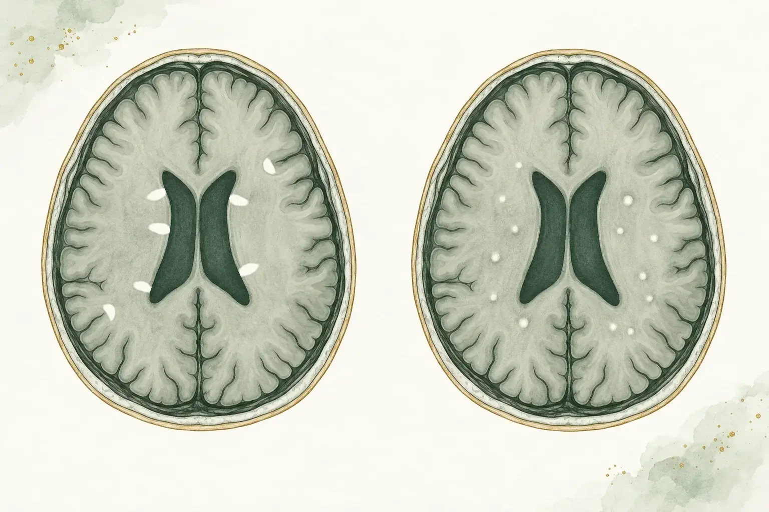
Two patterns of white spots
- The MS pattern (left): Oval spots sit against the fluid-filled ventricles, often pointing away from them, with others at the edge of the cortex.
- The small vessel pattern (right): Small, round dots are scattered in the deep white matter, away from the ventricle walls and the cortex.
- Read with the rest of the picture: The pattern is weighed with your age, symptoms and examination, and sometimes a spinal cord MRI or a follow-up scan.
White spots by age: your 20s to 40s, and after 60
Your age changes what a few white spots mean more than almost anything else on the report.
In your 20s to 40s. A few tiny spots are common in healthy people. In one study of 60 adults with no neurological symptoms, in their mid-30s on average, two in three had at least one. Migraine is a common reason at this age, and a far more common one than MS. A neurologist looks further when the spots are oval, touch the ventricles, sit at the edge of the cortex or in the brainstem or spinal cord, or come with symptoms that fit MS, such as blurred vision in one eye or numbness that spreads over days.
In your 40s and 50s. Blood vessel risk factors start to show. High blood pressure, diabetes, high cholesterol and smoking leave their first small marks, so a few dots become more common, and they are a reason to check and treat those risks.
At 60 and over. Most people have some. In the Rotterdam Scan Study of 1,077 people aged 60 to 90, only 1 in 20 had none. At this age the question is not whether spots are there, but how much change there is (the grade) and whether it grows on later scans.
Types of White Matter Lesions
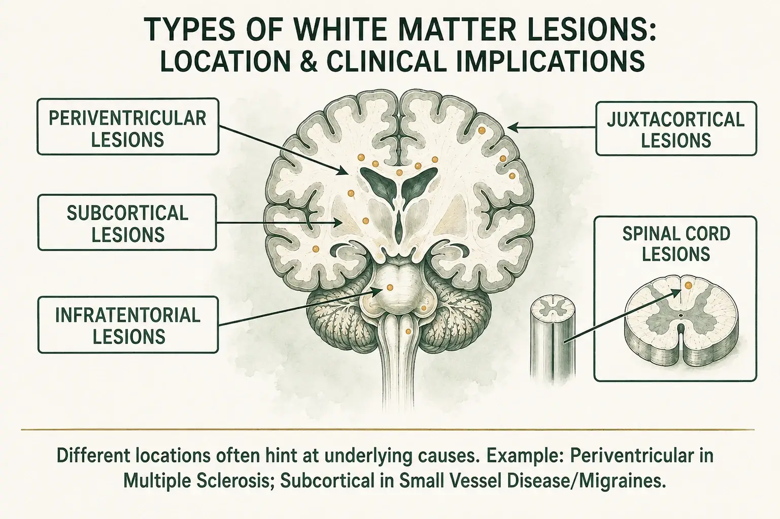
Reports name spots by where they sit. Here is what each location word means and what it usually points to:
| Report word | Where it is | What it usually points to |
|---|---|---|
| Supratentorial | Above the tentorium, a sheet of tissue over the cerebellum: the two cerebral hemispheres, almost the whole brain | A location word, not a diagnosis. Most spots of every cause sit here |
| Periventricular | Against the ventricles, the fluid-filled spaces in the middle of the brain | Smooth caps and thin bands are common with age. Oval spots that touch the ventricles and point away from them suggest MS |
| Juxtacortical | Touching the cortex, the brain's outer layer | One of the places MS favors |
| Subcortical or deep white matter | The white matter between the cortex and the ventricles, touching neither | Small round dots here are typical of small vessel change and migraine |
| Centrum semiovale, corona radiata | Names for the deep white matter above and beside the ventricles | The same as deep white matter: small dots here usually mean small vessel change |
| Periatrial | Beside the back corners of the ventricles | Small, faint, symmetric bright areas here can be a normal finding |
| Frontal or parietal lobe | The front of the brain, and the part behind it toward the top | A location only. Spots of every cause appear in these large lobes, so the shape and pattern say more than the lobe |
| Infratentorial | Below the tentorium: the brainstem and cerebellum | One of the places MS favors. With age and high blood pressure, small vessel change can also reach the middle of the brainstem |
| Spinal cord | The cord in the neck and back | Points toward an inflammatory cause such as MS or transverse myelitis. Small vessel change rarely shows here |
Location is a clue, not a verdict. The shape and number of the spots, the spinal cord and your symptoms count too, and many conditions can mimic MS on MRI.
Symptoms Associated with White Matter Lesions
Many individuals with white matter lesions might have no noticeable symptoms. However, depending on the severity and location, possible symptoms can include:
- Memory problems
- Difficulty with balance and coordination
- Slower cognitive processing, in MS, this is known as MS brain fog
- Mood changes, including depression or irritability
- Urinary urgency or incontinence (in severe cases)
- Vision changes or dizziness (especially in inflammatory conditions)
These symptoms can vary widely based on the cause and severity of the underlying issue. Note that muscle twitching or fasciculations, another symptom patients commonly worry about, are NOT typical features of white matter lesions; they usually point to peripheral nerve or muscle conditions rather than central brain lesions.
How Are White Matter Lesions Diagnosed?
The primary method for diagnosing white matter lesions is through neurological evaluation and magnetic resonance imaging (MRI). This non-invasive imaging technique provides detailed pictures of the brain and spinal cord, allowing neurologists to identify and assess white matter changes.
To diagnose white matter lesions, a neurologist usually reviews your MRI images to assess (see our guide on understanding your MS brain MRI for a detailed walkthrough):
- Size, number, and exact location of lesions
- Patterns that help distinguish between potential causes
- Changes over time through follow-up imaging
Additional tests, such as blood work, including newer biomarkers like neurofilament light chain (NfL), cognitive testing, spinal fluid analysis, or genetic testing, might be performed to identify specific underlying causes.
Three questions resolve most reports that say "nonspecific white matter changes." First, are any spots in the places MS favors, with a fitting shape: against the ventricles, at the edge of the cortex, in the brainstem or cerebellum, in the spinal cord, or, since the 2024 criteria, in the optic nerve? Small round dots in the white matter between the ventricles and the cortex, touching neither, are more typical of small vessel change or migraine. Second, was the spinal cord scanned, and was it clear? A report cannot describe what was never imaged. Third, do your symptoms, examination, age, and vascular risk factors fit?
No typical spots, a clear cord, and no clinical fit usually means "nonspecific" is the right word, and if you have vascular risk factors the spots are a reason to look after them. MS-typical spots found by chance in someone who has never had symptoms have a name, radiologically isolated syndrome, and a first episode of symptoms with fitting spots is a clinically isolated syndrome. A yes to any question, or a cord that was never scanned, means the images deserve a specialist read, not just the report. That last step matters: in a multicenter study of 110 people misdiagnosed with MS, "overreliance on MRI abnormalities in patients with nonspecific neurologic symptoms" was named as a leading contributor, and migraine was the most common correct diagnosis.
What Your MRI Actually Reveals
Your MRI doesn't just show "spots" - it provides critical information about lesion size, location, and pattern that helps distinguish between multiple sclerosis, vascular disease, migraines, and other conditions. Neurologists use the updated 2024 McDonald criteria to diagnose MS, which relies heavily on specific MRI patterns. This precise mapping guides your diagnosis and treatment strategy.
Treatment Options for White Matter Lesions
Managing white matter lesions involves addressing underlying causes and risk factors. Treatment strategies typically include:
Treat the cause, not the wording
- Context: The cause and clinical picture determine what needs treatment.
- Vascular health: Blood pressure and other risk factors may need attention.
- Follow-up: Further evaluation or imaging is based on the individual findings.
Managing Vascular Risk Factors
Since white matter lesions are often linked to vascular health, managing cardiovascular risk factors is a critical part of treatment. Here are some strategies that can help:
- Blood Pressure Control: Medications and lifestyle changes to lower blood pressure are key in preventing further brain tissue damage.
- Cholesterol Management: Statins and other medications, along with dietary changes, can help reduce the risk of white matter lesions.
- Blood Sugar Management: Keeping blood sugar levels in check if you have diabetes can slow the development of these lesions.
- Healthy Lifestyle: Regular exercise, a balanced anti-inflammatory diet, and avoiding smoking can all help protect your brain health.
Disease-specific Treatments
- Multiple Sclerosis and inflammatory disorders: Steroids (especially for acute relapses), immunotherapies, or disease-modifying drugs.
- Migraine Management: Preventive medications and lifestyle adjustments.
- Vitamin deficiencies: Nutritional supplementation (e.g., Vitamin B12).
Physical Therapy and Cognitive Support
For patients experiencing motor or cognitive symptoms, physical therapy may help improve balance and coordination. Occupational therapy can also help individuals adapt to daily tasks and maintain independence.
If cognitive symptoms such as memory loss or slowed thinking are present, neuropsychological testing, a 60-minute computerized test taken at home, measures what has changed, and cognitive rehabilitation strategies can help improve function.
Beyond Medication: Keys to Brain Protection
While medications target specific conditions, research shows that combining them with lifestyle changes can improve their effectiveness. Quitting smoking, Mediterranean diet adoption, and just 150 minutes of weekly exercise can have a significant impact on your brain health.
When to See a Neurologist for White Matter Lesions
A report full of "white matter hyperintensities" can keep you up at night, and most of the time the explanation is far less frightening than the words. In a 60-minute first visit for a multiple sclerosis evaluation, Dr. Ntranos reads the images with you, scan by scan, examines you, and tells you plainly what the spots mean and whether anything needs follow-up. If it does, follow-ups are usually every one to three months and last 30 to 45 minutes, and messages between visits are answered within 1 business day. Visits are in person in Beverly Hills or Los Angeles, or by video anywhere in California. Request a visit; no referral is needed, and the office confirms within 1 business day.
Living with White Matter Lesions
For many people, white matter lesions don't significantly impact daily life. However, proactive care can make a big difference:
- Follow your neurologist's recommendations closely.
- Maintain regular follow-up visits to track your brain health.
- Report new or worsening symptoms promptly.
- Adopt a healthy, active lifestyle to protect your brain.
White Matter Lesions and Flying
Pilots with white matter lesions on brain MRI may require an FAA neurological evaluation for medical certification. We provide comprehensive evaluations and documentation for pilots with neurological findings.
White Matter Lesions and Driving
Patients with white matter lesions may need a DMV driver evaluation to maintain their driving privileges. We provide comprehensive evaluations and documentation for patients with neurological findings.
Conclusion
White matter lesions are a complex topic, but understanding them is an important step in managing your brain health. While they can be concerning, many people with white matter lesions lead normal, healthy lives. By working closely with a neurologist who reads your images and focusing on overall health, you can take proactive steps to support your brain function and well-being.
If you have questions about white matter lesions or want a specialist to look at your MRI, our multiple sclerosis clinic in Los Angeles evaluates white matter findings, in person or by video.
Frequently Asked Questions About White Matter Lesions
References
- Wardlaw JM, et al. Neuroimaging standards for research into small vessel disease and its contribution to ageing and neurodegeneration. The Lancet Neurology, 2013.
- de Leeuw FE, et al. Prevalence of cerebral white matter lesions in elderly people: a population based magnetic resonance imaging study. The Rotterdam Scan Study. Journal of Neurology, Neurosurgery and Psychiatry, 2001.
- Palm-Meinders IH, et al. Structural brain changes in migraine. JAMA, 2012.
- Debette S, Markus HS. The clinical importance of white matter hyperintensities on brain magnetic resonance imaging: systematic review and meta-analysis. BMJ, 2010.
- Solomon AJ, et al. The contemporary spectrum of multiple sclerosis misdiagnosis: a multicenter study. Neurology, 2016.
- Nasrallah IM, et al. Association of intensive vs standard blood pressure control with cerebral white matter lesions. JAMA, 2019.
- Klawiter EC. Current and new directions in MRI in multiple sclerosis. Continuum, 2013.
- Keřkovský M, et al. Structural and functional MRI correlates of T2 hyperintensities of brain white matter in young neurologically asymptomatic adults. European Radiology, 2019.