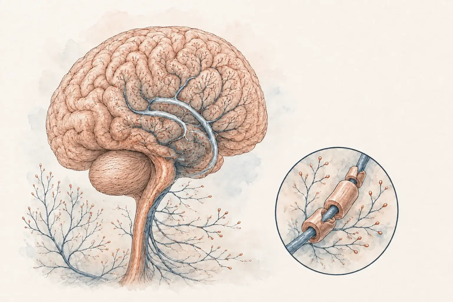
People arrive at an MS evaluation from two directions. Some have a symptom, such as blurred vision or numbness that climbed up a leg. Others have an MRI report with the word "demyelination" on it and a referral they did not expect. The evaluation answers whether this is MS or something else, and records a baseline to measure change against later. It starts with a 60-minute visit, and most people have an answer within a few weeks.
Dr. Achillefs Ntranos, MD is a board-certified neurologist who completed a fellowship in multiple sclerosis and demyelinating diseases at Mount Sinai and postdoctoral MS research at Johns Hopkins. Our Multiple Sclerosis Clinic page covers care after a diagnosis. This page is about reaching one.
What MS is, briefly
Multiple sclerosis is an immune-mediated disease in which inflammation damages myelin, the insulation around nerve fibers in the brain, spinal cord, and optic nerves. Where the damage lands determines the symptom. An optic nerve lesion dims vision in one eye, a spinal cord lesion causes numbness or weakness below it, a brainstem lesion causes double vision or vertigo.
The pattern that raises suspicion is an early symptom that develops over days, lasts weeks, and improves, in a younger adult. Fatigue and brain fog are common but point to many things.
What each test contributes
There is no single test for MS. The diagnosis is assembled from several pieces of evidence, each answering its own question.
| Test | What it contributes |
|---|---|
| History and neurological examination | Establishes that the symptoms are neurological and suggests where the lesions are |
| MRI of brain and spinal cord, with and without contrast | The most important test. Number, location, shape, and age of lesions |
| Blood tests | Cannot diagnose MS. They exclude the mimics: B12 deficiency, thyroid disease, infection, lupus, NMOSD, MOGAD |
| Spinal fluid, by lumbar puncture | Oligoclonal bands and kappa free light chains show the immune system is active inside the nervous system |
| Visual evoked potentials, optical coherence tomography | Evidence of old optic nerve damage that never caused a symptom |
Two things follow. A normal blood test does not rule MS out, and white spots on an MRI are not enough on their own, because migraine, small vessel disease, and several inflammatory conditions produce them too. See white matter lesions and MS or something else?.
Visual guide
Putting an MS evaluation together

- Clinical picture: the history and examination identify the symptom pattern.
- Imaging: brain and spinal cord MRI show the pattern of lesions.
- Other tests: blood tests investigate mimics; spinal fluid can add evidence when needed.
How the evaluation runs
The first visit, 60 minutes
Most of it is history. We go through every neurological episode you can remember, including ones years ago that resolved and were never explained. A bout of blurred vision at 24 can be the second episode the criteria require. The examination looks for signs that outlast symptoms: a subtle weakness, brisk reflexes, an eye that moves a fraction slower than its partner. The visit also sets your baseline: a timed walk and peg test in the exam room, and a computerized cognitive assessment you take at home, explained below. Send records ahead through our patient tools page.
Imaging and blood work, usually one to two weeks
An MS-protocol MRI of the brain, usually with the cervical and thoracic spinal cord, with and without contrast, at an imaging center near you and through your insurance. We handle the prior authorization. Dr. Ntranos reads the images himself and goes through them with you on screen rather than relaying the report. Our guide to reading an MS brain MRI explains the wording.
Spinal fluid, if the MRI does not settle it
We do not perform lumbar punctures in our office. We arrange it at a facility that does them under fluoroscopy, and the fluid goes to Mayo Clinic Laboratories. Results usually take one to two weeks.
The results visit, 30 to 45 minutes
In person or by video. What the evidence supports, what it does not, and either a diagnosis or the one test that would settle it.
The McDonald criteria
The McDonald criteria are the rules neurologists worldwide use to decide whether the evidence adds up to MS. They rest on three ideas.
- Damage in more than one place. Lesions in the MS-typical locations: around the ventricles, at the cortex, in the brainstem or cerebellum, in the spinal cord, and, since 2024, the optic nerve.
- Evidence of an ongoing process, not a single event. Historically that meant a second episode, new lesions on a later scan, or a mix of old and actively inflamed lesions on one scan. The 2024 revision no longer always requires it. When lesions sit in four or five of the typical locations, or when highly specific markers are present (oligoclonal bands or kappa free light chains in the spinal fluid, the central vein sign or paramagnetic rim lesions on advanced MRI), MS can be diagnosed at the first evaluation.
- No better explanation. Have the mimics been excluded? This rule did not change, and it is where most misdiagnoses happen.
Sometimes the honest answer after the first round of testing is that this may be MS and a repeat MRI after an interval will tell. We would rather say that than start long-term treatment on an uncertain diagnosis.
If a new symptom is developing now
New vision loss, weakness, numbness, or trouble walking that has come on over hours to days may be a relapse, whether or not you already have a diagnosis. Contact us the same day, and go to an emergency department if it is severe or worsening quickly. Our guide on what to do during an MS relapse has the steps.
Visual guide
What the results visit needs to answer

- Fit: do the symptoms, examination and imaging support MS?
- Alternatives: have conditions that mimic MS been considered?
- Uncertainty: repeat imaging or another test may be needed when evidence is incomplete.
Your baseline, measured at the first visit
Reaching a diagnosis is one job of the evaluation. Setting a starting point is the other. In the large relapsing MS trials, most disability accumulated slowly, without a relapse and often without a new lesion on MRI, and a change of that kind can only be seen against a baseline. So the first visit also records three standardized measures, and our Multiple Sclerosis Clinic repeats them.
- Timed 25-Foot Walk. Walk 25 feet as quickly and safely as you can, timed, twice. One number for legs, balance, and stamina. Repeated at every visit.
- Nine-Hole Peg Test. Nine pegs into nine holes and out again, each hand, timed. It tracks the fine hand function that typing, buttons, and cooking depend on. Repeated at every visit.
- Creyos cognitive assessment. A computerized neuropsychological battery rather than a five-minute screen: twelve tasks covering memory, attention, reasoning, planning, and processing speed, scored against norms for your age, plus validated mood, anxiety, and sleep questionnaires, because low mood and poor sleep lower scores in ways that mimic disease. About 60 minutes on your own computer at home. Repeated yearly, or sooner if something changes.
The walk and the peg test take about five minutes together. A 20% slowing on either, or a drop in a cognitive domain, that is still there months later counts as progression even when the MRI is unchanged. The walk and the peg test are the measures the trials use to define progression independent of relapse activity, explained in our guide to MS progression without relapses. The cognitive side is covered in MS brain fog and on our neuropsychological testing page.
After the evaluation, and requesting one
If it is MS, the next visit is about treatment. Disease-modifying therapies reduce relapses and new lesions, and the choice depends on how active your MS is, your other health conditions, and your family plans. Our MS treatment overview, the comparison of ocrelizumab, ofatumumab, and ublituximab, and the first five steps cover what we discuss. If it is not MS, you still leave with an answer: what the MRI findings mean, what caused the symptoms, and whether anything needs follow-up.
Being told it "might be MS" makes every week feel long. Request an evaluation if a neurological episode lasted more than a day, if an MRI report mentions demyelination, or if you want an existing diagnosis confirmed before treatment starts. The first visit is 60 minutes, in person in Beverly Hills or Los Angeles, or by video anywhere in California when your imaging already exists, and you leave understanding what has been found and what comes next. If you start by video, we say which baseline measures need an office visit. Follow-ups are usually every one to three months and last 30 to 45 minutes. Appointments are usually same or next business day, no referral is needed, and the office confirms your request within 1 business day. Visits are one flat fee with no facility charges, you receive a superbill, and most PPO plans reimburse 50–80%, depending on out-of-network benefits and deductible. See our billing page.
Frequently asked questions
References
- Montalban X, et al. Diagnosis of multiple sclerosis: 2024 revisions of the McDonald criteria. The Lancet Neurology, 2025.
- National Multiple Sclerosis Society. Diagnosing MS.
- National Multiple Sclerosis Society. Who gets MS.
- National Institute of Neurological Disorders and Stroke. Multiple sclerosis.
- Motl RW, et al. Validity of the timed 25-foot walk as an ambulatory performance outcome measure for multiple sclerosis. Multiple Sclerosis Journal, 2017.
- Feys P, et al. The Nine-Hole Peg Test as a manual dexterity performance measure for multiple sclerosis. Multiple Sclerosis Journal, 2017.
- Hampshire A, et al. Fractionating human intelligence. Neuron, 2012. The twelve-task battery behind the Creyos assessment.
- Kalb R, et al. Recommendations for cognitive screening and management in multiple sclerosis care. Multiple Sclerosis Journal, 2018.