
If your feet burn at night, or your hands tingle and sting, and a nerve conduction study came back normal, someone has probably mentioned a skin biopsy. The natural question is whether a test on a tiny piece of skin can really settle it. Accurate enough to rely on, with one limit: the biopsy finds small fiber neuropathy in about 8 of every 10 people who have it, so a normal result makes the condition less likely but does not rule it out. The procedure itself is covered in our guide to what to expect during a small fiber skin punch biopsy.
Understanding skin biopsy accuracy: sensitivity and specificity
Two numbers describe how good any medical test is.
- Sensitivity is how often the test is positive in people who truly have the condition. A sensitivity of 80 percent means it catches 80 of every 100 people with small fiber neuropathy and misses 20.
- Specificity is how often the test is negative in people who do not have it. A specificity of 90 percent means 90 of every 100 people without the condition get a correctly normal result, and 10 get a false alarm.
For skin biopsy measuring intraepidermal nerve fiber density (IENFD), across published series, sensitivity is roughly 75 to 90 percent and specificity roughly 65 to 90 percent, depending on the cohort and the laboratory's normal values. Those are strong figures for a nerve test. They are not perfect, and the gap matters most on the sensitivity side: about one person in five with genuine small fiber neuropathy can have a biopsy that reads as normal.
The study that set the diagnostic criteria makes the comparison concrete. Of 486 people screened for suspected small fiber neuropathy, 67 had the pure form, with normal nerve conduction studies. In that group skin biopsy had a diagnostic efficiency of 88%, against 55% for the clinical examination and 47% for quantitative sensory testing. The biopsy does not replace the examination, and the criteria combine the two. But it was the single most informative test, by a wide margin.
The measurement itself is simple in principle. A 3 mm punch of skin is taken from the lower leg, stained with a marker called PGP9.5 that lights up every nerve fiber, and the fibers crossing into the outer skin layer are counted per millimeter. Your count is compared with reference values for your age and sex. A count below the fifth percentile is abnormal.
Why skin biopsy is the gold standard for SFN diagnosis
Skin biopsy earned its place as the reference test for four reasons.
- It looks at the fibers directly. Every other small fiber test measures function, such as how you perceive temperature or how much you sweat. The biopsy counts the fibers themselves, which removes most of the subjectivity.
- It sees what EMG cannot. Nerve conduction studies and EMG test large, insulated fibers. Small fibers are too thin and too slow to register, which is why a normal EMG is expected in this condition rather than reassuring.
- It uses standardized reference ranges. International guidelines set the biopsy site, the stain, the counting rules, and the age- and sex-matched normal values, so a result from a good laboratory means the same thing in Los Angeles as in Milan.
- It gives a number you can repeat. A fiber density can be compared with a later biopsy to see whether the neuropathy is stable, improving, or progressing.
What the guidelines say about skin biopsy and autonomic testing
Patients often search for the American Academy of Neurology position on this, so here is the short version. The joint guideline of the European Federation of Neurological Societies and the Peripheral Nerve Society, first published in 2005 and updated in 2010, recommends skin biopsy with PGP9.5 staining as the objective test of choice for small fiber neuropathy and sets the technical standards that laboratories follow. The American Academy of Neurology's 2009 practice parameter on distal symmetric polyneuropathy, since retired, described skin biopsy as a validated technique for measuring nerve fiber density that may be considered for diagnosis, particularly of small fiber sensory neuropathy.
The same documents treat autonomic tests, quantitative sensory testing, and corneal confocal microscopy as supporting rather than primary tests. In practice, a neurologist combines the biopsy with the history and examination, and adds autonomic testing when symptoms such as light-headedness on standing, abnormal sweating, or digestive changes suggest the autonomic small fibers are involved.
Why a normal EMG does not rule out small fiber neuropathy
This is the single most common misunderstanding we see. A patient with burning feet has an EMG and nerve conduction study, is told everything is normal, and concludes the pain must be coming from somewhere else. It usually is not.
EMG and nerve conduction studies measure the large myelinated fibers that carry strength, vibration sense, and position sense. Small fibers carry pain and temperature and run the autonomic nervous system. They are unmyelinated or thinly myelinated, and standard electrodes simply cannot record from them. A normal EMG therefore tells you that the large fibers are fine. It says nothing about the small ones. When symptoms sound like small fiber neuropathy and the EMG is normal, the skin biopsy is the next test, not the end of the road. Our neuropathy in the feet guide explains how the two fiber types produce different symptoms.
Reading your result: positive, borderline, or normal
A positive result means the fiber count is below the fifth percentile for your age and sex. Combined with typical symptoms and a normal EMG, that is diagnostic of small fiber neuropathy. It does not tell you the cause, which becomes the next question.
A borderline result means the count sits close to the cutoff. Laboratories report these honestly rather than forcing them into one box. A borderline biopsy in someone with clear symptoms is often treated as supportive, and a repeat biopsy after a year can show whether the count is falling.
A normal result lowers the likelihood of small fiber neuropathy but does not remove it, for the reasons covered below. A normal biopsy in someone with strong symptoms is a reason to look again at the pattern of symptoms, consider a non-length-dependent form, and sometimes add autonomic testing.
Whatever the number, it is read alongside your story and examination. An abnormal count in someone with no symptoms is not a diagnosis, and a normal count in someone with classic symptoms is not a dismissal.

Read the result alongside the symptoms
- Low fiber count: A reduced count can support small fiber neuropathy in the right clinical setting.
- Borderline result: A count near the cutoff needs careful interpretation.
- Normal result: A normal biopsy lowers the likelihood but does not completely exclude it.
The terms on your report
Skin biopsy reports use a handful of technical terms. This is what each one means in plain words.
| On your report | What it means |
|---|---|
| Intraepidermal nerve fiber density (IENFD, or ENFD) | The number of small nerve fibers that cross into the outer layer of the skin, counted per millimeter of the sample |
| Normal range, or 5th percentile for age and sex | The laboratory's cutoff for people of your age and sex. A count below the fifth percentile is abnormal |
| Distal leg and proximal thigh | The two sample sites: just above the outer ankle (distal, farther from the trunk) and the upper outer thigh (proximal, closer to it) |
| Length-dependent or non-length-dependent | Whether the loss is greater at the ankle than at the thigh, the common pattern that starts in the feet, or patchy, with the thigh as low as the ankle or lower |
| Sweat gland nerve fiber density | A separate count of the autonomic fibers wrapped around the sweat glands, the nerves that control sweating |
| Axonal swellings | Swollen points along the fibers. Some studies link them to early damage, but the fiber count carries more weight |
| Congo red | An extra stain that shows amyloid deposits, added when amyloidosis is a question |
Your report prints the laboratory's own normal range next to each count. Those ranges depend on age, sex and the laboratory itself, so a number from one report should not be judged against a cutoff from another.
Skin biopsy for autonomic neuropathy
The same skin sample contains sweat glands, and the same PGP9.5 stain shows the tiny autonomic fibers that wrap around them. These sudomotor fibers are lost in autonomic neuropathy, the condition behind symptoms such as heat intolerance, patchy or absent sweating, light-headedness on standing, and digestive or bladder changes described on our autonomic dysfunction page.
Studies of sweat gland innervation report that skin biopsy is abnormal in 96 percent of patients when the lower leg is sampled and 79 percent at the thigh, in a series of 28 patients, which outperformed routine autonomic testing. Loss of these fibers is not seen in people whose neuropathy is purely sensory, so the finding helps separate the two. It also correlates with symptoms: the fewer sweat gland fibers, the more likely someone is to report impaired sweating and heat intolerance.
QSART, the quantitative sudomotor axon reflex test, measures sweat output directly and has a sensitivity of roughly 50 to 80 percent for distal small fiber neuropathy. It needs specialized equipment that few centers have, which is why the biopsy, which answers the same question from the same sample, is often the more practical route.
Amyloid detection: a special capability
Amyloidosis is a group of conditions in which misfolded protein builds up in tissues, including nerves. Two types cause neuropathy: hereditary transthyretin amyloidosis (ATTRv), which runs in families, and light chain (AL) amyloidosis, which is linked to plasma cell disorders. Both matter because effective treatments now exist, and both work best when started early.
When amyloidosis is a possibility, the skin sample can be stained with Congo red, which binds amyloid deposits. Reported sensitivity varies widely between series, from roughly 50 percent to near 100 percent, and is highest when both the ankle and thigh samples are examined with many sections. Specificity is very high: amyloid was found in none of 58 controls in the largest skin-biopsy series. In plain terms, a positive Congo red stain is close to certain, while a negative one does not fully exclude the disease. The amount of amyloid found also tracks with the degree of nerve fiber loss, which gives some sense of how advanced the process is. If there is a family history of neuropathy with heart or kidney involvement, or unexplained weight loss and carpal tunnel on both sides, it is worth asking whether the stain should be added.
Factors that affect skin biopsy accuracy
Where and how the sample is taken. The standard site is 10 cm above the outer ankle bone, chosen because length-dependent neuropathies show up there first. A sample taken from the wrong site, or too shallowly, can under-count fibers.
Which laboratory reads it. This is the biggest variable. As the JAMA review puts it, normative values differ between laboratories, so the same biopsy can be interpreted differently in different places. Laboratories that specialize in nerve fiber counting, publish their own reference ranges, and run quality control produce more reliable results. Our biopsies are analyzed by Therapath, a laboratory dedicated to this work.
How far along the neuropathy is. Sensitivity is highest in established, moderate disease, where fiber loss is easy to see. In very early or mild disease the count may not yet have dropped below the cutoff, even though the fibers are already misbehaving.
Other medical conditions. Diabetes tends to increase the yield of the test, because diabetic small fiber damage is usually clear. Autoimmune causes more often produce a patchy, non-length-dependent pattern that a single leg sample can miss. Vitamin B12 deficiency and thyroid disease can cause similar symptoms and are checked with blood tests alongside the biopsy.
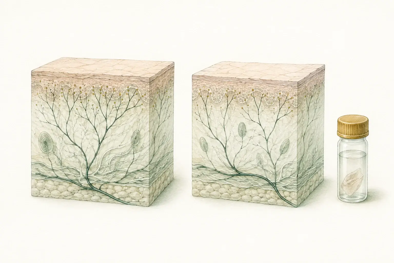
Can a skin biopsy miss small fiber neuropathy?
Yes, in a minority of cases. The recognized reasons are:
- Very early disease. Symptoms can appear before enough fibers are lost to cross the threshold.
- Patchy involvement. If the damage is scattered rather than starting at the feet, the sampled site may be spared.
- Non-length-dependent neuropathy. Some forms, often autoimmune, affect the trunk, face, or arms first, and a leg biopsy can be normal.
- Borderline counts. A count just above the cutoff is technically normal but not reassuring.
- Fibers that are present but not working. Some medication effects and metabolic conditions disturb small fiber function without yet reducing fiber number.
A follow-up study of people with suspected small fiber neuropathy found that those with both an abnormal biopsy and abnormal sensory testing at the start were the most likely to develop clear clinical signs later, while those with only one abnormal test rarely progressed. That is a useful way to think about a normal biopsy: it is one strong piece of evidence, and it is weighed with the others rather than ending the discussion.
Why a test can miss a condition
- Sampling location: The sampled area may not represent every symptomatic location.
- Disease pattern: Patchy or non-length-dependent involvement can complicate interpretation.
- Follow-up: Persistent symptoms may warrant another look at the clinical picture.
Comparing skin biopsy to other SFN tests
| Test | What it measures | Sensitivity | Specificity | Notes |
|---|---|---|---|---|
| Skin biopsy (IENFD) | Number of small nerve fibers in the skin | 75–90% | 65–90% | Reference test; objective; repeatable |
| Quantitative sensory testing (QST) | Thresholds for temperature and vibration | 47–78% | 39–70% | Depends on attention and effort |
| QSART | Sweat gland output | 50–80% | Moderate | Few centers offer it |
| Corneal confocal microscopy | Small fibers in the cornea | Variable | Variable | Research use mostly; not widely available |
| EMG and nerve conduction | Large fiber function | Not applicable | Not applicable | Normal in pure small fiber neuropathy |
Head-to-head studies consistently find that skin biopsy outperforms quantitative sensory testing, which has a subjective component and wide variability. The most reliable picture comes from combining the examination, one or more functional tests, and the biopsy.
What happens after the result
A confirmed diagnosis changes two things. First, it starts a search for the cause: blood sugar and glucose tolerance, vitamin B12, thyroid function, autoimmune markers, and a protein screen for monoclonal gammopathy, with genetic or amyloid testing when the history points that way. Some of those causes can be treated and the neuropathy stabilized or partly reversed, which is the subject of our article on whether peripheral neuropathy can be reversed. The cause is also what shapes what small fiber neuropathy means for life expectancy. Second, it gives you and your neurologist an objective baseline, which makes future decisions about symptom treatment and monitoring easier to justify.
Hearing that your EMG is normal while the burning or tingling goes on is confusing, and it is not the end of the road. Dr. Ntranos sees you first for a 60-minute visit, in person in Beverly Hills or Los Angeles, or by video anywhere in California. If a biopsy would answer the question, he performs it in our Beverly Hills office after that first evaluation. The report usually takes about two weeks, sometimes three when sweat gland density is included, and we go through it with you alongside your examination and blood work, so you understand what the number means. Follow-ups are usually every one to three months and last 30 minutes, and messages between visits are answered within 1 business day. Request a visit; no referral is needed, and the office confirms within 1 business day. Details are on our page about small fiber neuropathy testing in Beverly Hills.
Frequently asked questions
References
- Peripheral Neuropathy: A Review. JAMA, 2025.
- Scientific Advances in and Clinical Approaches to Small-Fiber Polyneuropathy: A Review. JAMA Neurology, 2019.
- European Federation of Neurological Societies / Peripheral Nerve Society guideline on the use of skin biopsy in the diagnosis of small fiber neuropathy. European Journal of Neurology, 2010.
- The Diagnostic Challenge of Small Fibre Neuropathy: Clinical Presentations, Evaluations, and Causes. Lancet Neurology, 2017.
- Skin Biopsy in the Management of Peripheral Neuropathy. Lancet Neurology, 2007.
- Which Method for Diagnosing Small Fiber Neuropathy? Frontiers in Neurology, 2020.
- Diagnosis of small fiber neuropathy: a comparative study of five neurophysiological tests. Neurophysiologie Clinique, 2015.
- Peripheral autonomic neuropathy: diagnostic contribution of skin biopsy. Journal of Neuropathology and Experimental Neurology, 2012.
- The diagnostic criteria for small fibre neuropathy: from symptoms to neuropathology. Brain, 2008.
- American Academy of Neurology. Practice Parameter: Evaluation of Distal Symmetric Polyneuropathy, Role of Autonomic Testing, Nerve Biopsy, and Skin Biopsy. Neurology, 2009.
- Diagnostic accuracy of intraepidermal nerve fiber density in small fiber neuropathy. PubMed Central, 2020.
- Autonomic testing: how to interpret and when to order. Cleveland Clinic Journal of Medicine, 2018.
- Diagnostic yield of skin biopsy for amyloid deposits in transthyretin and light chain amyloidosis. Neurology, 2024.