If your neurologist has suggested a skin biopsy for small fiber neuropathy, the word biopsy may sound bigger than the procedure is. This is a skin sample the size of a pencil tip, not surgery on a nerve. A numbing injection stings for a few seconds, each sample takes seconds, there are no stitches, and you can drive yourself home.
What is a small fiber skin punch biopsy?
A skin biopsy for neuropathy removes two tiny round samples of skin, 3 mm across, from the leg. Each sample contains the endings of the small nerve fibers that carry pain and temperature. A specialized laboratory stains those fibers, counts them under a microscope, and compares the count to normal values for your age and sex. A low count confirms small fiber neuropathy.
The test exists because the small fibers are invisible to nerve conduction studies and EMG, which measure only the large fibers. If you have burning, stabbing, or tingling feet and a normal EMG, this is the test that can still give you an answer. It is called a punch biopsy because the tool is a small hollow cylinder that removes a round core of skin, like a very small cookie cutter.
It is sometimes confused with a nerve biopsy, which surgically removes a piece of a nerve. That is a different, more involved procedure that is rarely needed. The skin biopsy is what is used for small fiber neuropathy. How well it performs, and what a normal result can and cannot rule out, is covered in how accurate is a skin biopsy for small fiber neuropathy.

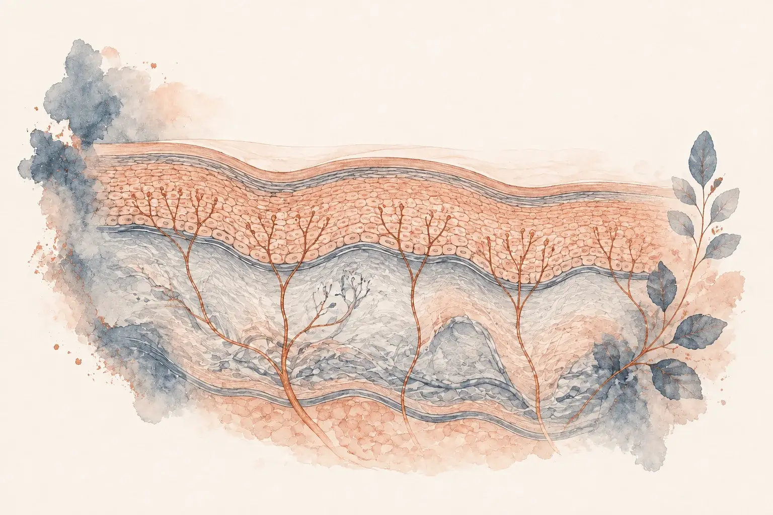
What a skin biopsy looks for
Epidermis
The outer skin layer. Small sensory nerve endings reach into this layer from the dermis below.
Small nerve fibers
These fine endings help you sense pain and temperature.
Skin sample
The laboratory stains the sample and counts nerve fibers, comparing their density with reference values.
Before your skin punch biopsy
The biopsy is scheduled after a neurological evaluation, where Dr. Ntranos examines you and decides whether the test will answer your question. If your own doctor suggested the test, request a visit directly: no referral is needed, the first visit can be by video anywhere in California, and most people come to our Beverly Hills office once, for the biopsy itself. Small fiber neuropathy testing in Beverly Hills covers the laboratory, the timing and billing.
Blood work for treatable causes of neuropathy is usually ordered at that first visit, so the two results can be read together.
There is no preparation. Eat and drink as usual and keep taking your medications. If you take a blood thinner, tell us when we schedule the biopsy so we can plan for it; in most cases nothing changes. Wear or bring shorts, or trousers that roll easily above the knee, since the samples come from the outer ankle and the upper outer thigh of one leg. You do not need anyone to drive you.
During the skin punch biopsy procedure
Dr. Ntranos performs the biopsy himself in our Beverly Hills office. You lie on the examination table with one leg exposed. Two sites are cleaned and marked: one just above the outer ankle and one on the upper outer thigh. Two sites are used so the fiber count near the foot can be compared with the count higher up, which tells us about the pattern of the neuropathy and not just whether it is present.
Each site gets a small injection of local anesthetic. This is the part that stings, for a few seconds, much like a dental injection. Within a minute or two the skin is numb. The 3 mm punch is then pressed and turned to take the sample, which takes seconds. You may feel pressure or a slight tug, but not sharp pain. Light pressure stops the small amount of bleeding, and a small adhesive bandage goes on. No stitches are needed for a wound this size.
The samples go into preservative and ship to the laboratory the same day. Most people say the whole thing was easier than they expected.

A small sample, a laboratory answer
- Local numbing: The skin is numbed before each small sample is taken.
- Two sites: Samples from near the ankle and upper thigh help show the pattern.
- Aftercare and results: Follow the written wound-care instructions while the laboratory processes the samples.
After your skin punch biopsy
You can drive yourself home and go back to work or your usual day. The sites feel like a small scrape for a day or two. You leave with written aftercare instructions covering bandage changes, showering, and when to avoid soaking the sites, and you can call us with any question in the meantime. Contact us sooner if a site becomes increasingly red, warm, swollen, or starts to leak, since that can mean a minor infection that is easy to treat.
How long does a punch biopsy take to heal?
A 3 mm punch site closes on its own in about a week to ten days. This is what it usually looks like along the way:
| When | What the site looks like |
|---|---|
| The first day or two | Tender, like a small scrape |
| About a week | A small scab |
| 7 to 10 days | Closed, once the scab falls away |
| The following months | A small pale dot that slowly fades |
| A year later | Hard to find; most people cannot see the sites |
Vigorous exercise and swimming wait until the sites have closed; the written instructions cover the specifics.
Getting your results
Your samples are analyzed at Therapath, a laboratory that specializes in nerve fiber counting. The report gives an epidermal nerve fiber density, meaning the number of small fibers per millimeter of skin, at each site, alongside the normal range for your age and sex.
Results usually take about two weeks, sometimes three when sweat gland density is included, and we tell you when yours is back. At the results visit Dr. Ntranos goes through the numbers with you. A reduced count at the ankle with a normal count at the thigh is the commonest pattern and indicates a length-dependent small fiber neuropathy. A reduced count at both sites suggests a different process, often an autoimmune one, which changes the search for a cause. A normal count does not exclude every form of small fiber neuropathy, and the accuracy article explains why.
The biopsy tells us the fibers are reduced, not why. What comes next, from finding and treating the cause to managing the pain, is covered on neuropathy in the feet and can peripheral neuropathy be reversed.
Burning or tingling feet and a normal EMG is a frustrating combination, because that test cannot see the small fibers that carry pain. With or without that EMG, a 60-minute first visit with Dr. Achillefs Ntranos shows whether the biopsy is the right next step for you, and why. It can be in person in Beverly Hills or Los Angeles, or by video anywhere in California; the biopsy itself is done in the Beverly Hills office. Follow-ups are usually every one to three months and last 30 minutes, and messages between visits are answered within 1 business day. Request a visit; no referral is needed, and the office confirms within 1 business day.

From laboratory result to the next step
- Laboratory analysis: The laboratory stains and counts the small nerve fibers.
- Clinical interpretation: The count is compared with reference information and your symptoms.
- Next steps: The result helps guide further evaluation and a treatment discussion.
Frequently asked questions
References
- Lauria G, et al. European Federation of Neurological Societies/Peripheral Nerve Society Guideline on the use of skin biopsy in the diagnosis of small fiber neuropathy. European Journal of Neurology, 2010.
- National Institute of Neurological Disorders and Stroke. Peripheral Neuropathy.
- Cleveland Clinic. Small Fiber Neuropathy.
- Therapath Neuropathology. Epidermal Nerve Fiber Density.
- Therapath Neuropathology. Skin Biopsy: specimen handling and turnaround.
- Johns Hopkins Medicine. Cutaneous Nerve Laboratory.