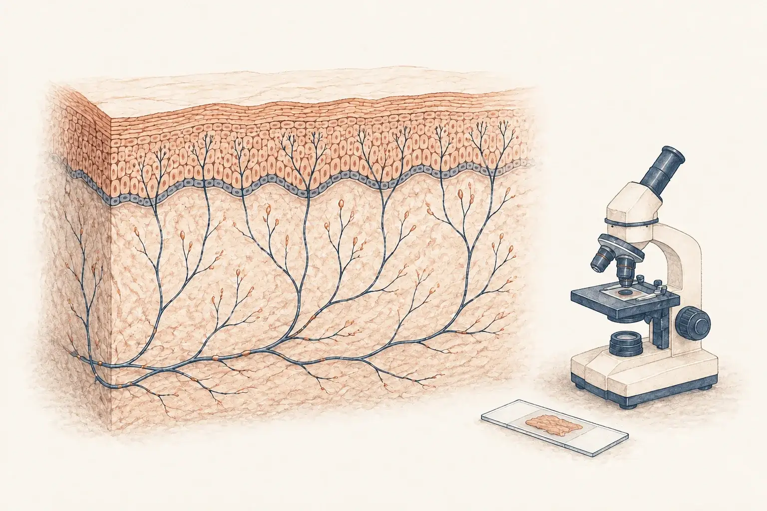
If you have burning pain in your feet and a normal EMG (the standard electrical nerve test), the nerves that carry pain were never actually tested. A skin biopsy tests them directly, by counting the nerve endings in a sample of skin the size of a pencil eraser.
Who should get small fiber neuropathy testing
The typical patient we biopsy has had burning pain in the feet for months, a normal EMG, and has been told the nerves are fine. EMG measures the large myelinated fibers. It cannot see the small ones that carry pain, so a normal result leaves small fiber neuropathy untested.
- Burning, electric, or pins-and-needles pain in the feet. Our neuropathy in the feet guide describes the pattern.
- Pain from light touch, such as socks or bed sheets, often worse at night.
- Reduced pain or temperature sensation with normal strength and balance.
- Autonomic symptoms: abnormal sweating, heat intolerance, lightheadedness on standing.
- A background of diabetes or prediabetes, an autoimmune condition, or long COVID.
It is not the first test for everyone with numb feet. Weakness or poor balance points to the large fibers, where nerve conduction studies come first. Numbness in one hand is more often carpal tunnel syndrome or a pinched nerve.
What small fiber neuropathy is
Small fiber neuropathy is damage to the thinnest nerve fibers, the ones carrying pain and temperature and running the autonomic nervous system. The trunks still conduct, which is why standard testing passes. What is lost is the fine terminal branches in the skin.
Symptoms usually begin in the toes and climb in a stocking-and-glove pattern, reaching the hands later. A minority have a patchy, length-independent pattern instead, which is more often autoimmune.
Because the large fibers are intact, the neurological examination is often normal too: strength, reflexes, and balance all pass, and the only clue may be reduced pinprick or temperature sensation in the feet. A normal examination does not make the pain imaginary. It makes the biopsy the right test.
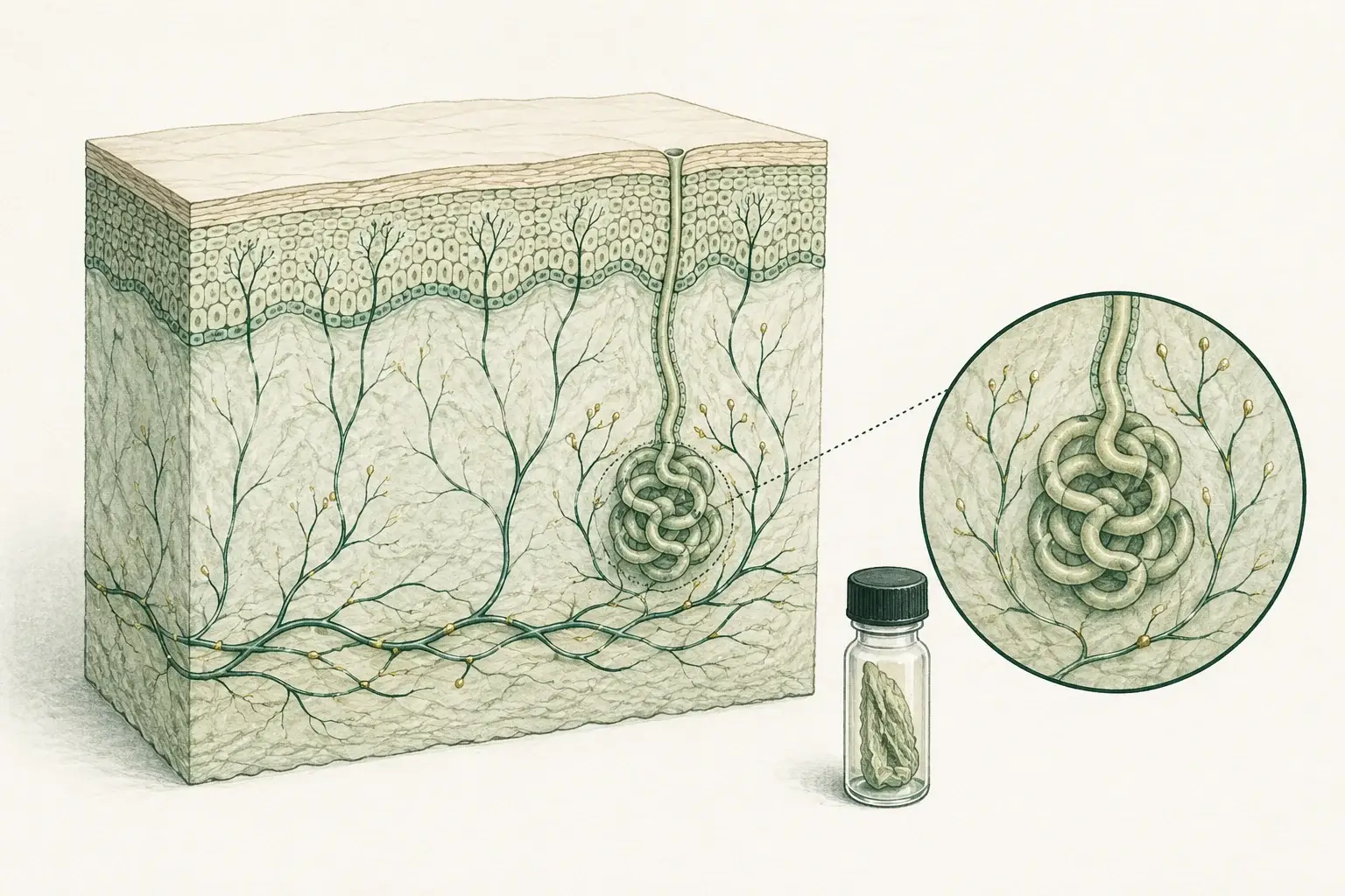
The nerve endings a skin biopsy examines
- Sensory fibers: fine endings carry pain and temperature signals.
- Autonomic fibers: fibers around sweat glands help assess autonomic involvement.
- Different test: EMG and nerve conduction studies assess the larger fibers.
Which test diagnoses small fiber neuropathy
| Test | What it answers |
|---|---|
| Skin punch biopsy | Counts the small fibers directly against normal values. The reference test, built for the patient whose EMG was clean |
| EMG and nerve conduction studies | Large fibers only. Normal in pure small fiber neuropathy, useful for a mixed picture. We refer for these |
| Autonomic function testing | Sweating, heart rate, and blood pressure responses. Done at centers we refer to |
| Blood tests | Not the diagnosis, the cause: glucose tolerance, B12, thyroid, celiac and autoimmune markers |
The biopsy gives a number rather than a rating of what you felt, so it can be repeated later. Its limits are worth hearing from us: a normal count does not exclude every form, technique and normative data affect the result, and it tells you the fibers are reduced, not why.

What a skin biopsy looks for
Epidermis
The outer skin layer. Small sensory nerve endings reach into this layer from the dermis below.
Small nerve fibers
These fine endings help you sense pain and temperature.
Skin sample
The laboratory stains the sample and counts nerve fibers, comparing their density with reference values.
The biopsy, step by step
A visit first
Sixty minutes with Dr. Achillefs Ntranos, MD, in person or by video, to decide whether the biopsy answers your question. Blood work for a treatable cause is ordered then.
On the day
No fasting. Take your usual medications, tell us about blood thinners, and wear loose pants.
The punch
Two sites are marked, the outer ankle and the upper outer thigh, so the two can be compared. Each is numbed with a small injection, the part that stings. A 3 mm punch takes each sample in seconds, and the appointment at our Beverly Hills office takes 45 to 60 minutes.
Afterwards
A small adhesive bandage, no stitches. You leave with written aftercare instructions, and most people return to normal activity the same day.
Results
Samples ship to Therapath the same day. The report usually takes about two weeks, sometimes three when sweat gland density is included, and we tell you when yours is back and book a follow-up to go through it.
Two articles go further: what to expect at the biopsy, and how accurate skin biopsy is.
Your result, and what it means
| Finding | What it means |
|---|---|
| Low at the ankle, normal at the thigh | Length-dependent small fiber neuropathy, the commonest form |
| Low at both sites | A length-independent process, more often autoimmune, which changes the search for a cause |
| Sweat gland fiber density low | Autonomic fibers are involved, which affects the workup and the treatment |
At the results visit you get the numbers, a diagnosis or a working diagnosis, and a written plan.
Autonomic fibers from the same samples
The stain also shows the fibers wrapped around each sweat gland, so one procedure answers two questions. That matters if you have abnormal sweating, heat intolerance, fainting on standing, a diagnosis of POTS, or dry eyes and mouth. Sweat gland density can be low even when the epidermal count is normal, so we request it every time.
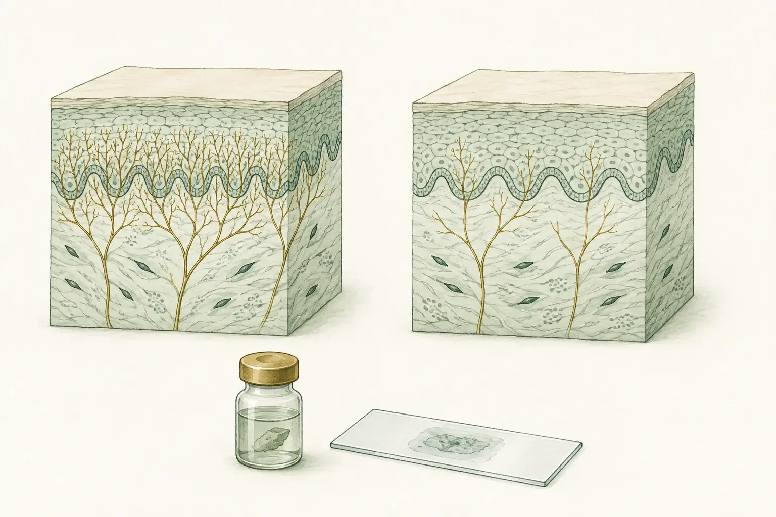
What a fiber count can tell you
- Density: the biopsy measures small fibers in the sampled skin.
- Pattern: ankle, thigh and sweat-gland findings help guide the workup.
- Limits: reduced fibers do not explain the cause, and a normal count does not exclude every form.
What happens after the diagnosis
- Treat the cause. Blood sugar control, replacing vitamin deficiencies, thyroid treatment, and immune therapy through our autoimmune neurology service. Prediabetes is a common, reversible driver.
- Control the pain. Several medication classes act on nerve pain rather than ordinary pain, chosen around your other conditions.
- Daily life. Exercise, foot care, sleep, and limiting alcohol measurably affect nerve pain. Our article on whether neuropathy can be reversed sets out what is realistic.
In a fair share of patients no cause is found. Symptom control then becomes the main work, and a repeat biopsy shows whether things are stable.
Small fiber neuropathy testing in Beverly Hills and Los Angeles
Dr. Ntranos, a board-certified neurologist, performs every biopsy himself at our Beverly Hills office. Consultations and results visits can also be at our West Los Angeles office or by video, so patients who travel come in once. We do not perform EMG or autonomic testing here, and refer for both.
Visits and the biopsy are flat-fee and not billed to insurance. You receive a superbill, and most PPO plans reimburse 50–80%, depending on out-of-network benefits and deductible. The laboratory bills separately. See our billing page.
Your small fiber neuropathy specialist
Dr. Ntranos is a board-certified, fellowship-trained neurologist with research training in neuroimmunology at Johns Hopkins. That matters here, because some of the treatable causes of small fiber neuropathy are autoimmune. One doctor sees you through every step: he examines you, takes the samples, reads the report with you, and treats both the cause and the pain. Follow-ups are usually every one to three months, and messages between visits are answered within 1 business day.
Burning feet and a normal EMG can leave you feeling dismissed. Request a visit or call (310) 774-7025; no referral is needed, and the office confirms your request within 1 business day. Have a result from elsewhere? Request a second opinion.
Frequently asked questions
References
- Lauria G, et al. European Federation of Neurological Societies/Peripheral Nerve Society guideline on the use of skin biopsy in the diagnosis of small fiber neuropathy. Eur J Neurol. 2010;17(7):903-912.
- Practical Neurology. Diagnosing small fiber neuropathy through the use of skin biopsy.
- National Institute of Neurological Disorders and Stroke. Peripheral neuropathy.
- Therapath Neuropathology. Epidermal nerve fiber density.
- Therapath Neuropathology. Skin biopsy: specimen handling and turnaround.
- Johns Hopkins Medicine. Cutaneous Nerve Laboratory.