
Transverse myelitis usually arrives without warning: numbness climbing from the feet, legs that feel heavy, a tight band around the chest, trouble emptying the bladder, all over a few days. Once the emergency is over and the name has been given, most people run into the same question. Does this mean I have multiple sclerosis?
Sometimes, and often not. Transverse myelitis can be a single event with no sequel, or it can be the first attack of MS or of a related condition such as NMOSD or MOGAD. Which of those is true decides your long-term treatment, and it is a question that can be answered with the right tests. This guide explains what transverse myelitis is, how neurologists tell it apart from MS, what recovery tends to look like, and what to ask for.
What is transverse myelitis?
Transverse myelitis is inflammation of a segment of the spinal cord. "Myelitis" means inflammation of the cord; "transverse" describes inflammation that extends across its width, though in practice many cases involve only part of the cross-section. The inflammation damages myelin, the insulating coating around nerve fibers, and sometimes the fibers themselves, so signals between the brain and the body below that level are disrupted.
The spinal cord carries every command going down to the muscles and every sensation coming up from the body. A lesion in the thoracic cord (mid-back) typically affects the legs, trunk, bladder, and bowel. A lesion in the cervical cord (neck) can affect the arms as well.
Transverse myelitis is a description of what is happening, not a final diagnosis. It has several possible causes: an autoimmune disease of the central nervous system such as MS, NMOSD, or MOGAD; an infection or the immune response that follows one; a systemic autoimmune condition such as lupus or sarcoidosis; or no identifiable trigger, which is called idiopathic transverse myelitis. Finding the cause is the whole point of the evaluation, because MS, NMOSD, and MOGAD each need different long-term treatment, and a medication that helps MS can worsen NMOSD.

Inflammation within the spinal cord
- Where it happens: Inflammation affects a segment of the spinal cord.
- What can change: Weakness, altered sensation, and bladder or bowel symptoms may occur.
- Why testing matters: MRI, spinal fluid, and blood tests help determine the underlying cause.
Early signs and symptoms of transverse myelitis
Symptoms typically build over hours to days, occasionally over one to two weeks. The pattern is neurological trouble below a particular level of the spinal cord.
- Weakness. Heaviness or clumsiness in the legs, sometimes progressing to difficulty walking or, in severe cases, paralysis.
- Numbness or tingling. Reduced or altered sensation, often starting in the feet and rising to a definable level on the trunk.
- A band-like sensation. A squeezing or tight feeling around the chest or abdomen, sometimes called the "MS hug". This is often one of the earliest signs.
- Pain. Back pain at the level of the inflammation, or burning and shooting pain in the legs or trunk.
- Bladder and bowel changes. Urgency, difficulty emptying, incontinence, or constipation.
- Sensitivity to temperature and touch. Ordinary sensations feeling unpleasant or distorted.
Severity varies enormously, from mild numbness and a slightly unsteady walk to complete loss of movement below the waist within hours. The speed and pattern of onset are themselves clues: a partial, one-sided picture that evolves over days is more typical of MS, while rapid, severe, symmetrical involvement raises the question of NMOSD.
Symptoms that need emergency care
Rapidly worsening weakness, new inability to urinate, loss of bowel control, or numbness spreading upward over hours should be evaluated in an emergency department the same day. Spinal cord inflammation is treated most effectively when it is treated early, and the same symptoms can also come from spinal cord compression, which is a surgical emergency.
Transverse myelitis vs MS: how neurologists tell them apart
MS is one of the most common causes of transverse myelitis, and a spinal cord attack is a frequent first presentation of MS. But transverse myelitis and MS are not the same thing. MS is a chronic disease that produces repeated episodes of neuroinflammation across the brain, spinal cord, and optic nerves over years. Transverse myelitis is a single episode confined to the cord. The question is whether that episode is the first attack of MS, the first attack of something else, or a one-time event.
Several features help.
- The spinal cord lesion itself. MS lesions are usually short, spanning one or two vertebral segments, and sit toward the edge of the cord, often on one side. Lesions that extend over three or more segments and fill the center of the cord are more typical of NMOSD or MOGAD.
- Partial versus complete. MS tends to cause partial myelitis, with symptoms affecting one side more than the other or sensation more than strength. Complete transverse myelitis, with severe symmetric loss of movement and sensation, points away from MS.
- The brain MRI. White matter lesions in MS-typical locations at the time of the spinal cord attack are the strongest single predictor that this is MS.
- Spinal fluid. Oligoclonal bands, a marker of immune activity inside the central nervous system, are found in roughly nine out of ten people with MS and are less common in NMOSD and MOGAD.
- Antibody blood tests. Aquaporin-4 antibodies identify NMOSD, and MOG antibodies identify MOGAD. Both can be checked from a blood sample.
Put together, these findings usually sort a spinal cord attack into one of three groups: MS or likely MS, an antibody-mediated condition, or idiopathic myelitis to be monitored.
Can transverse myelitis turn into MS?
It can, and the risk is not the same for everyone. When a first attack is accompanied by MS-typical lesions on brain MRI, most people go on to develop MS. The best long-term data come from a study that followed people after a first attack of any type, whether optic neuritis, a brainstem episode, or a spinal cord attack, and found that about 80 percent had developed MS 20 years later. When the brain MRI is normal, the figure is closer to one in five. Positive oligoclonal bands, a short partial cord lesion, and a younger age at onset all raise the likelihood further.
The picture is different for idiopathic transverse myelitis, meaning a spinal cord attack with a normal brain MRI, negative antibody tests, and no other cause found. In a large series of such patients, about 13 percent were diagnosed with MS over roughly three years of follow-up, and most never had another episode.
This is why the evaluation matters so much. Under the 2024 McDonald criteria, MS can now be diagnosed at the time of a first attack if the MRI and spinal fluid findings meet the criteria, and treatment can begin without waiting for a second episode. If the criteria are not met, the plan is monitoring with repeat MRI, so that a change is caught early. Until then, the first attack is called a clinically isolated syndrome.
How transverse myelitis is diagnosed
The evaluation has two goals: confirm inflammation of the cord, and find its cause. A thorough workup usually includes the following.
- Spinal cord MRI with contrast. This is the central test. It shows where the lesion is, how many segments it spans, whether it sits at the edge or the center of the cord, and whether it enhances with contrast, which indicates active inflammation. It also excludes compression from a disc or a mass, which can produce similar symptoms and needs completely different treatment.
- Brain MRI. Essential even when all the symptoms are in the spinal cord, because the presence and pattern of brain lesions is the strongest guide to whether this is MS. Our guide to understanding your MS brain MRI explains what the report is describing.
- Spinal fluid analysis. A lumbar puncture, which we arrange with interventional radiology, provides fluid that is checked for oligoclonal bands, white blood cells, and protein, and for infection when the history suggests it.
- Blood tests. Aquaporin-4 and MOG antibodies; markers of systemic autoimmune disease such as lupus, Sjögren's, and sarcoidosis; vitamin B12 and copper, which can cause spinal cord problems that mimic myelitis; and infection screens where relevant.
Occasionally a spinal cord attack looks like textbook MS and the antibody tests reveal NMOSD instead. That single result changes the long-term treatment entirely, which is why the full panel is worth running at the first episode rather than assuming the most common explanation.
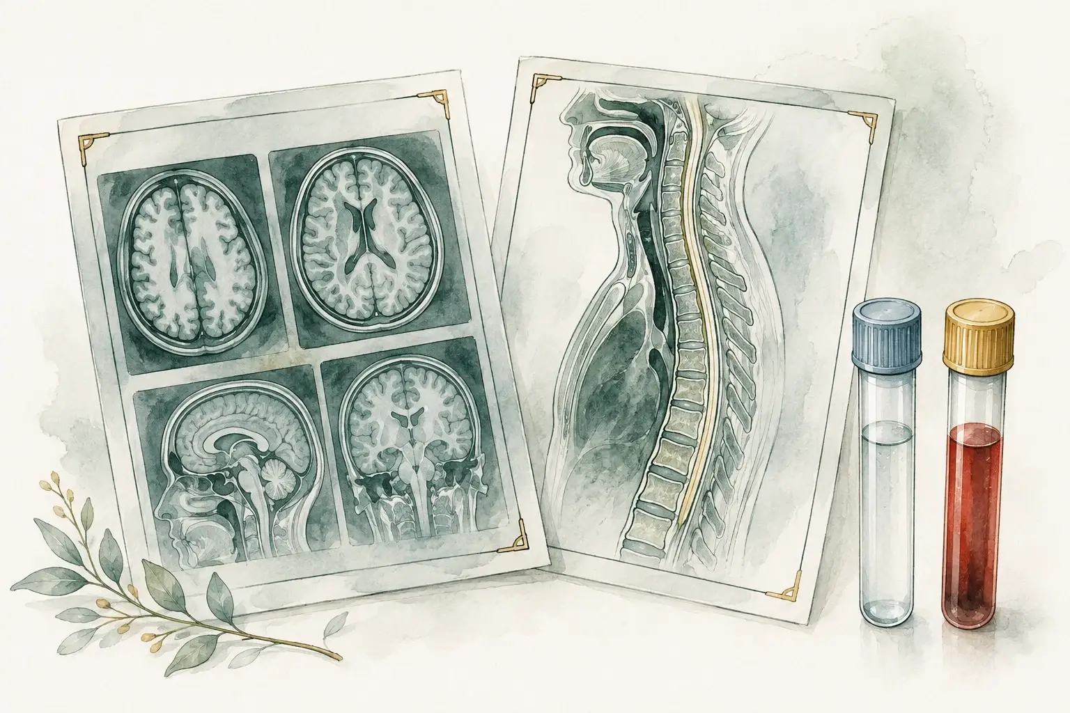
Testing helps explain the spinal inflammation
- Imaging: Brain and spinal cord MRI show the location and pattern of inflammation.
- Spinal fluid: Fluid testing can add evidence about inflammation.
- Antibody tests: Blood testing helps assess conditions such as NMOSD and MOGAD.
What transverse myelitis looks like on MRI
The spinal cord MRI often points toward the cause before the antibody results come back. These are the usual patterns.
| Feature | MS | NMOSD | MOGAD | Idiopathic |
|---|---|---|---|---|
| Lesion length | Short, usually one or two vertebral segments | Long, three or more segments | Often long; can be short, and there may be more than one lesion | Varies |
| Position in the cord | Toward the edge, often on one side | The center, often most of the cord's width | The central gray matter, often reaching the lower tip of the cord (conus) | Varies |
| Brain MRI | MS-type lesions are common, even without brain symptoms | Often normal, or a few lesions in typical spots such as the brainstem | Often normal, or large, fluffy lesions unlike MS | No MS-type lesions, by definition |
| Contrast enhancement | A new lesion often enhances for a few weeks | Often enhances during the attack, sometimes in a patchy or ring pattern | Often little or none | Varies |
Scans do not always follow the pattern. An NMOSD lesion is sometimes short, and MS lesions can merge into a longer one, so the MRI is read together with the spinal fluid and antibody results.
Treatment: the acute attack and the long term
Treating the attack
The first priority is to reduce the inflammation quickly. The standard treatment is a short course of high-dose corticosteroids, usually given intravenously, with the form and length chosen by the treating neurologist. Steroids speed recovery, though they do not by themselves change the eventual outcome. For severe attacks that do not respond to steroids, plasma exchange, which filters antibodies and other immune components from the blood, is the next step, and it is particularly effective in NMOSD-related myelitis. Rehabilitation begins as soon as the acute phase settles.
Treatment after the attack
What happens next depends entirely on the cause.
- If this is the first attack of MS, starting a disease-modifying therapy substantially reduces the risk of further relapses and of long-term disability. Highly effective options, including the anti-CD20 therapies, are often considered after a spinal cord attack because cord relapses are more likely to leave lasting deficits.
- If this is NMOSD or MOGAD, different medications are used. Several MS therapies are ineffective in NMOSD and some can make it worse.
- If no cause is found, the plan is monitoring with repeat MRI over the following years. Many people never have another episode; a minority are later diagnosed with MS or another condition, and early detection is the reason for the follow-up.
Recovery time and outlook
How long transverse myelitis takes to recover is the question almost everyone asks, and the honest answer is a range. In most people improvement begins within a few weeks to three months of the attack. The fastest gains come in the first months, and slower improvement can continue for a year or two. Progress is rarely a straight line; plateaus followed by further gains are common.
A typical recovery timeline looks like this, though each attack sets its own pace.
| Time since the attack | What usually happens |
|---|---|
| First days | Symptoms reach their worst point, usually within days. Steroids, and plasma exchange if needed, calm the inflammation while MRI, spinal fluid and blood tests look for the cause. |
| Weeks 2 to 12 | Improvement usually begins. Rehabilitation starts as soon as the acute phase settles: physical and occupational therapy, plus bladder care when needed. |
| Months 3 to 12 | Recovery continues, often at its fastest early in this period. Repeat MRI checks for new lesions, especially when the cause is still unclear. |
| Years 1 to 2 | Slower improvement can continue. Care shifts to whatever remains, such as stiffness, nerve pain or bladder problems, and to preventing another attack when there is an underlying cause. |
Outcomes across all causes of transverse myelitis follow what neurologists call the rule of thirds: about a third recover with little or no lasting deficit, a third are left with moderate problems such as some weakness, numbness, or bladder symptoms, and a third have significant permanent disability. Several things shift where an individual is likely to land.
- Severity at the worst point. People who keep some movement and sensation at the nadir (the lowest point of the attack) recover better than those with complete paralysis.
- Speed of onset. Very rapid onset over hours tends to carry a worse outlook than evolution over days.
- Cause. MS-related partial myelitis generally recovers better than NMOSD-related attacks.
- Early treatment and rehabilitation. Prompt steroids and sustained physical, occupational, and bladder rehabilitation improve the chances of a good outcome.
Is transverse myelitis life-threatening?
Rarely. Transverse myelitis itself is not usually fatal, and it does not shorten life expectancy in most people. The exceptions are attacks high in the cervical cord that affect breathing, which are treated in hospital, and complications of severe disability such as infections or blood clots, which good long-term care is designed to prevent. Most of the questions about outlook are really about function, not survival, and function is where treatment and rehabilitation make their difference.
Can transverse myelitis come back?
Idiopathic transverse myelitis usually happens once. A second episode is a strong signal that an underlying condition such as MS, NMOSD, or MOGAD is present and should prompt a full re-evaluation. In people already diagnosed with one of those conditions, further attacks are exactly what long-term treatment is meant to prevent; our guide to MS relapses covers what to do if new symptoms appear.
When to see an MS specialist
After an episode of transverse myelitis, it is worth having the full picture reviewed by a neurologist who works with demyelinating diseases every day. That is especially true if your brain MRI shows lesions nobody has fully explained, or you were told you have "possible MS" and are unsure what comes next. It also applies if your antibody tests were never done or were unclear, if you are deciding about treatment, if recovery has stalled, or if you have had more than one episode. A multiple sclerosis evaluation reviews the actual MRI images, the spinal fluid and antibody results, and your examination together, and a second opinion is reasonable for a diagnosis that will shape years of treatment.
"Does this mean MS?" is the question most people carry home, and the right tests go a long way toward answering it. In a 60-minute first visit, Dr. Achillefs Ntranos goes through that evidence with you, so you know what it supports and what comes next. Follow-ups are usually every one to three months and last 30 to 45 minutes, and messages between visits are answered within 1 business day. Request a visit in Beverly Hills or Los Angeles, or by video anywhere in California. No referral is needed; the office confirms within 1 business day.
Frequently asked questions
References
- National Institute of Neurological Disorders and Stroke. Transverse myelitis.
- Cleveland Clinic. Transverse myelitis (TM): causes, symptoms and treatment.
- Fisniku LK, et al. Disability and T2 MRI lesions: a 20-year follow-up of patients with relapse onset of multiple sclerosis. Brain, 2008.
- Cobo-Calvo A, et al. Idiopathic acute transverse myelitis: outcome and conversion to multiple sclerosis in a large series. BMC Neurology, 2013.
- Dobson R, et al. Cerebrospinal fluid oligoclonal bands in multiple sclerosis and clinically isolated syndromes: a meta-analysis of prevalence, prognosis and effect of latitude. Journal of Neurology, Neurosurgery and Psychiatry, 2013.
- Montalban X, et al. Diagnosis of multiple sclerosis: 2024 revisions of the McDonald criteria. The Lancet Neurology, 2025.