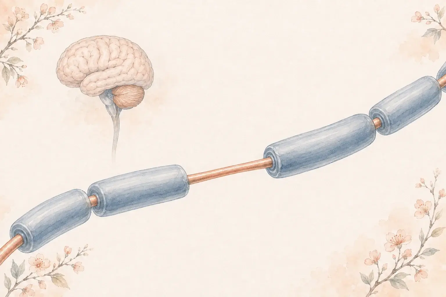
Clinically isolated syndrome, or CIS, is a first episode of symptoms such as blurred vision in one eye, numbness, or double vision, caused by inflammation that damages myelin, the insulation around nerve fibers. The episode usually improves over weeks, often almost completely. On its own it is not a diagnosis of multiple sclerosis, and some people never have another episode.
What clinically isolated syndrome is
Multiple sclerosis is diagnosed by showing that demyelination has happened in more than one place in the central nervous system and, in most cases, at more than one time. A first episode, by definition, can only show one time. Clinically isolated syndrome, or CIS, is the name for that first episode when it has the character of MS but the evidence does not yet add up to the diagnosis. It is a description of where you are in the process, not a separate disease.
The label is honest rather than evasive. Some people who have a single episode never have another, and their scans stay clean. Others are in the first chapter of relapsing-remitting MS. The work of the first visit is to tell those two groups apart as accurately as the evidence allows, and to set out a plan for whichever one you are in.

Where a first episode happens
- Optic nerve: Dim or blurred vision in one eye with pain on eye movement.
- Spinal cord: Numbness, weakness, or a band of tightness, often on one side or below one level.
- Brainstem: Double vision, vertigo, facial numbness, or unsteadiness.
Symptoms
A CIS episode has a shape. It comes on over hours to days, plateaus, lasts weeks, and then improves, often almost completely. There is no fever or infection to explain it. The three classic presentations:
- Optic neuritis. Vision in one eye becomes dim, blurred, or washed out over a few days, with an ache behind the eye that is worse on movement. Colors look faded. Most people recover most of their vision over weeks to months.
- Partial transverse myelitis. Numbness, tingling, or weakness that rises over days, often on one side or below a level on the trunk, sometimes with a tight band or bladder urgency. Our guide to transverse myelitis and MS covers this form in depth.
- Brainstem or cerebellar syndrome. Double vision, vertigo, facial numbness, slurred speech, or clumsiness and unsteadiness.
A CIS can also involve more than one of these areas at once, which points more strongly toward MS.
What are the chances it becomes MS?
The MRI at the time of the first episode is the strongest predictor we have. In the long-term studies, people whose first scan showed MS-typical white matter lesions had a high chance of a second attack or new lesions within a few years, and most had a diagnosis of MS within a decade. People whose first scan was clean had a low chance, on the order of one in five even after twenty years. The number of lesions, their location, and whether the spinal fluid shows oligoclonal bands or kappa free light chains, markers of immune activity inside the nervous system, refine that estimate further.
| First episode | First brain MRI | Chance of MS in the long-term studies |
|---|---|---|
| Any type | Normal | About 1 in 5 over 20 years |
| Any type | MS-typical lesions | About 4 in 5 over 20 years |
| Optic neuritis | Normal | About 25 in 100 at 15 years |
| Optic neuritis | One or more lesions | About 72 in 100 at 15 years |
The figures for any type of first episode come from a study that followed people for 20 years (reference 7); the optic neuritis figures come from the Optic Neuritis Treatment Trial (reference 8). Both began before today's treatments, so they largely describe the course without disease-modifying therapy, and your own odds depend on the details of your scan and spinal fluid.
The 2024 McDonald criteria changed what happens with that information. When a first episode comes with lesions in enough of the typical locations, or with specific spinal fluid or advanced MRI markers, MS can now be diagnosed at that first visit rather than waiting for a second event. The practical effect is that fewer people are left with the CIS label, and those who are have genuinely uncertain findings and a clear monitoring plan.
How CIS is evaluated
The MS evaluation after a first episode has two jobs. The first is to make sure the episode was demyelination and not one of the conditions that mimic MS: a stroke, a compressed nerve or spinal cord, a vitamin B12 deficiency, an infection, or an inflammatory disease elsewhere in the body. The second is to look for the other demyelinating diseases that present the same way and are treated differently. Optic neuritis and myelitis are also the classic first attacks of NMOSD and MOG antibody disease, so antibody blood tests for aquaporin-4 and MOG are part of the workup when the picture fits.
The core tests are MRI of the brain and full spinal cord with contrast, blood tests, and often a lumbar puncture. Optic neuritis may add an eye examination and optical coherence tomography, a painless scan of the retina.
Treatment and what happens next
The episode itself is usually treated with a short course of high-dose steroids when it is disabling, which speeds recovery. The longer question is whether to start a disease-modifying therapy. For people whose MRI shows MS-typical lesions, the trials are consistent: starting treatment after a first episode delays the second and reduces new lesions, and the benefit persists years later. For people with a clean scan, the risk is low enough that watching with a repeat MRI is reasonable. In between, the decision is made together, weighing the lesion burden, the spinal fluid, your age, plans for family, and your own view of risk. Our treatment guide explains the options.
Vitamin D has been tested at this stage too. In a 2025 trial of people with a first episode typical of MS who had not started treatment, high-dose vitamin D modestly reduced new activity, mostly on MRI. It does not replace a disease-modifying therapy, and the dose is set after a blood test; our guide to vitamin D and MS explains what the trials show.
Whatever is decided, the plan should be specific: when the next MRI is due, what change would confirm MS or prompt treatment, and what to do if a new symptom appears in the meantime.

After a first episode
- Repeat MRI: Scheduled months apart to look for new lesions, with or without symptoms.
- Decision point: New lesions or a second episode usually confirm MS and change treatment.
- Between scans: A new symptom lasting more than 24 hours is a reason to call, not wait.
What to ask after a first episode
A first episode leaves you with more questions than answers. These are the ones worth bringing to your neurologist:
- Does my MRI show lesions in the places typical of MS, and how many?
- Were my brain and my whole spinal cord scanned, with contrast?
- Would a lumbar puncture change the plan for me?
- Have NMOSD and MOG antibody disease been ruled out with blood tests?
- Do I already meet the 2024 McDonald criteria for MS?
- What would starting a disease-modifying therapy now do for me, and what are the trade-offs?
- When is my next MRI, and what result would change the plan?
- What should I do if a new symptom appears before then?
When to see a neurologist
After a first episode of blurred vision, numbness or double vision, one question hangs over everything: is this MS? Some people never have another. See a neurologist within days rather than weeks for vision loss in one eye with pain, numbness or weakness that builds over days, or double vision or vertigo that does not settle. Call 911 or go to an emergency department instead if symptoms start within minutes, vision is lost suddenly, weakness spreads over hours, or you cannot pass urine. Once the episode has been evaluated, an MS specialist is the right person to interpret the MRI and decide with you whether to treat. At our Multiple Sclerosis Clinic, Dr. Ntranos, a fellowship-trained MS specialist, reviews your images directly in a 60-minute first visit, usually available the same or next day, and you leave knowing when the next MRI is due and what would change the plan. MRI and any lumbar puncture are arranged at outside facilities. Follow-ups are usually every one to three months and last 30 to 45 minutes, and messages between visits are answered within 1 business day. Visits are in Beverly Hills or Los Angeles, or by video anywhere in California; request a visit without a referral, and the office confirms within 1 business day.
Frequently asked questions
References
- Miller DH, et al. Clinically isolated syndromes. The Lancet Neurology, 2012.
- Kappos L, et al. The 11-year long-term follow-up study from the randomized BENEFIT CIS trial. Neurology, 2016.
- Montalban X, et al. Diagnosis of multiple sclerosis: 2024 revisions of the McDonald criteria. The Lancet Neurology, 2025.
- National Institute of Neurological Disorders and Stroke. Multiple sclerosis.
- National Multiple Sclerosis Society. Types of MS.
- Thouvenot E, et al. High-Dose Vitamin D in Clinically Isolated Syndrome Typical of Multiple Sclerosis: The D-Lay MS Randomized Clinical Trial. JAMA, 2025.
- Fisniku LK, et al. Disability and T2 MRI lesions: a 20-year follow-up of patients with relapse onset of multiple sclerosis. Brain, 2008.
- Optic Neuritis Study Group. Multiple sclerosis risk after optic neuritis: final optic neuritis treatment trial follow-up. Archives of Neurology, 2008.